A herniated disc is a common and painful condition affecting hundreds of thousands of Americans every year.
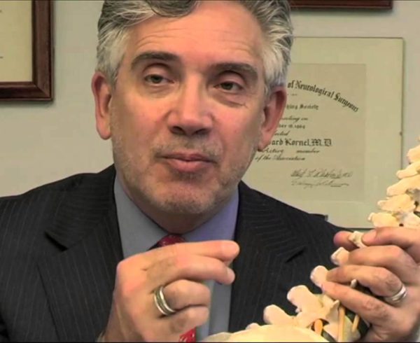
Noted New York Neurosurgeon, Dr. Ezriel Kornel is a specialist in minimally invasive neurological and spinal procedures and is in the forefront in bringing relief to patients suffering from the pain of a herniated disc. Dr. Kornel is a partner in Brain and Spine Surgeons of New York and has offices in Manhattan and White Plains. He is an Assistant Clinical Professor of Neurosurgery at Weill Cornell Medical College in New York and is affiliated with several hospitals in Westchester County and Manhattan.
Dr. Kornel explains that a herniated disc occurs when a disc extrudes from the confines of the disc itself to compress a nerve in the spine. As the spinal canal has nerves running through it, a portion of a degenerated disc is extruded into the spinal canal, compressing the nerve and causing pain.
The lumbar spine is located in the lower back. The spinous process is the bone you can feel when you run your hand up and down along your back. “They flare out to form the roof of the spine, known as the lamina,” Dr. Kornel explains. “Further out, they form the joints of the spine, and allow for movement of the spine. Inside the spinal canal runs the nerve, which we refer to as the cauda equina, or horse’s tail. In the front of the spine we have the vertebral bodies, which are the building blocks of the spine.” In between are the ‘cushions,’ which are the discs. “The nerves come out or goes into the spine through the opening called the spinal foramen between the discs and the joints. When a disc herniates it can compress the nerve as it’s coming out, or it can compress multiple nerves within the spinal canal,” he says.
Removal of just an extruded fragment of the disc is what Dr. Kornel refers to as a micro discectomy. “Or, the way I perform the procedure is referred to as an endoscopic micro discectomy.” The standard approach, which was used for many years, was to enter the spine through an incision in the center of the spine. There are muscles, which come across to attach to the spine that pull the spine, allowing for movement. There are ligaments between the vertebrae that hold the vertebrae together and the joints together.
“In order to get into the spine in the standard technique, the muscles and the tendons that hold the muscles to the bone have to be cut, and the muscles are pulled over to expose the bone,” Dr. Kornel explains. “A portion of the bone is then removed, which is referred to as a laminectomy.” (That is also the generic term for any spine surgery that involves removal of a disc.) “However,” he adds, “we don’t remove the entire lamina, and most times even with the most standard techniques only a portion of the lamina is removed. That allows for access into the spine. Once in the spine, the nerves can be moved aside and the extruded disc fragment is able to be plucked out.
However, Dr. Kornel often elects to perform the procedure minimally invasively. “With a micro discectomy, the incision is a bit smaller,” he explains. “In order to visualize the nerves well and the extruded disc well, a microscope is used to magnify the operative site. The technique that I like to use, which is a minimally invasive technique, is what is referred to as an endoscopic microdiscectomy. It means that rather than dividing the muscles and tendons off of the spine, we’re able to separate out the muscle fibers through a very small — about two centimeters — opening in the skin. And then we insert a tube through the skin, separating the muscle down to the edge of the bone. Through that tube we can then insert an endoscope with a fiber-optic camera and see the internal structure that way, or we can actually look through the tube with a microscope, which is the way I prefer to do it; that gives us a direct, three-dimensional image.
“Then we can enter the space between the vertebrae, between the lamina,” Dr. Kornel continues. “Often because the lamina are overlapping we have to remove a small amount of it, but generally we remove a small portion, about the size of a fingernail, to enable us to gain access. There is a ligament, called the yellow ligament, that goes between the lamina and overlies the nerve. That ligament helps to protect the nerve. Through this approach we need to remove very little of that ligament. I generally move only a small, small portion of that ligament — only a few fibers — off to the side to gain access into the spinal canal from the side.
“Once we gain access, we can see the nerve and we can see the extruded disc fragment because we have such high magnification we’re able to see these structures well,” Dr. Kornel explains. “And then, through that very small opening, we’re able to pluck out that fragment of disc, relieving the pressure on the nerve. We can then remove the tube, the muscles come back together, and we make a small closure in the skin. Usually we don’t have to use sutures on the outside — all the sutures are buried, so there are no stitches to remove, and patients go home the same day.”
Generally the pain from the herniated disc that has caused pressure on the nerve is relieved right away, and people feel the relief of pain when they wake up. “There is certainly some soreness to the back because there is an incision made — there’s no way to get into the spine without making an incision.
“My patients are walking around and go home the same day and they are able to be up and about,” he continues. “I like my patients to rest and to limit their physical activities for several weeks. The likelihood of that disc re-herniating is small.”
The likelihood of that disc herneating again in the future is somewhere around 15%, “especially if you take care of your back after the surgery,” Dr. Kornel explains, “meaning if you strengthen your core muscles, if you watch your alignment and if you’re careful about the way you use your back, that you don’t try to move heavy furniture without help, that you are attentive to the way you’re moving, the likelihood of the reherniation is quite small.
Dr. Kornel concludes, “the surgery is very effective in relieving the symptoms, and the likelihood that a patient will be able to live a normal, functional life is very high, well above 90%.”


